Multi-level mass spectrometric characterisation of Antibody-Drug Conjugates in a regulated environment

Eric Largy
Anicet Catrain
Arnaud Delobel
Analytical Technologies Europe 2017

Anti body-Drug Conjugates (ADCs) are anti bodies engineered to deliver a cytotoxic agent specifi cally to tumor cells expressing a defi ned anti gen.

The challenge of human mesenchymal stem cells (MSCs) characterisation

Sandra Thys, R&D Scientist

Access this 45 minute webinar to learn how to characterise human mesenchymal stem cells in terms of identity, purity and biological activity. The analytical performance of the methods developed have been discussed along with the presentation of experimental results obtained during method development, optimisation and/or validation.

Human mesenchymal stem cells are widely used in the field of cell therapy, a technology used for the replacement and regeneration of dysfunctional cells by healthy functional cells.

The characterisation of human MSCs used for manufacturing cell therapy products requires the development and validation of analytical methods which meet regulatory requirements. Cell therapy products emerge from a long process of cell culture, differentiation and purification where appropriate cell identity, purity and biological activity (potency) monitoring is mandatory. According to minimal criteria proposed by the International Society for Cellular Therapy (ISCT), MSCs must have the ability to adhere to cell culture plastic surfaces, express some phenotypic markers and differentiate into adipogenic, osteogenic and chondrogenic lineages.

Identity and purity tests can be based on the analysis of one or more specific markers by flow cytometry, fluorescent microscopy and/or qPCR. Biological activity (potency) can be measured using enzymatic assays or immunoassays linked to the relevant biological properties of the cell therapy product. We will present some efficient, optimised approaches developed at Quality Assistance for the characterisation of human mesenchymal stem cells

Cytokine Release: Comparison of Multiplex and Simplex assays

Quality Assistance

Access this 45 minute webinar to learn:

  • How to overcome the challenges usually associated to Multiplex assays
  • How to select the right technology for your needs

Cytokine profiling is a powerful tool to link the host immune system with disease pathogenesis and/or treatment efficacy. 
The analysis of cytokine expression is frequently required for the characterisation of the status of the immune system, to demonstrate the function of the cells of interest and as an analysis of surrogate markers for in vitro potency testing. 

Classically, the analysis of cytokine expression is performed by ELISA for each separate analyte. 
Multiplex assays allow the simultaneous measurement of multiple cytokines in a sample, increasing the efficiency (less time-consuming and less expensive) and considerably reducing the sample volumes needed.

In the context of a 9 month internal R&D project, we have compared three multiplex technologies for the measurement of cytokines:

  • Electrochemiluminescence (ECL, MSD)
  • Magnetic beads (Luminex, Merck Millipore) 
  • Cytometric Bead Array (FACS Verse, BD)
  • The performance of the three assays was evaluated in terms of their accuracy, repeatability, intermediate precision, quantification range and ability to detect cytokines in human PBMC samples. 

Customised labelling of antibodies: A powerful tool for multi-parameters analyses in Flow Cytometry

Valérie Deffontaine
Fabian Vandermeers
Capucine Lepers
Géry Van Vyncht
Arnaud Delobel

The characterisation of biomarkers by flow cytometry is an attractive approach for Quality Control applications of cell products. Unfortunately, the design of a customised panel remains challenging, especially for stem cell products. The aim of this study was to generate customised labelled antibodies with three classes of fluorochromes allowing multi-parameters analyses by flow cytometry. The “in-house” labelled antibodies were compared to their commercial counterparts and their stability was evaluated. These antibodies were used in the context of a method validation to evaluate their intrinsic performances.

Our protocol allows the labelling of antibodies with fluorochromes that match the customer’s and regulator’s needs. The “16/3” validation design used in this study allows the generation of a complete dataset suitable for meaningful statistical analyses.

Mass spectrometry toolkit for the characterisation of protein glycosylation

Quality Assistance

Access this 45 minute webinar to learn how state-of-the-art LC/MS technologies can be implemented for the efficient study of therapeutic protein glycosylation using analyses optimised for each level of information you need. 

Analytical performance of the different solutions proposed have been discussed, together with the presentation of numerous experimental results (with advantages and drawbacks) obtained during method development and optimisation. 

We presented efficient and optimized approaches for the characterisation of protein glycosylation by UPLC/mass spectrometry, based on different levels of analysis: 

  • Fast and highly sensitive profiling of released N-glycans, with use of the RapiFluor-MS® technology, 
  • Specific release and profiling of O-glycans,
  • Orthogonal released glycans analysis and sialylation profiling by mixed-mode chromatography, 
  • Unambiguous determination of N- and O-glycosylation sites by widepore-HILIC peptide mapping, 
  • High-resolution intact and subunit analysis. 

Applications of these methods to mAbs (Humira®, Erbitux®) and highly glycosylated proteins (Enbrel®) will be presented. 

The performances and limitations of the different analytical approaches will be discussed. 

Characterisation of the glycosylation of Humira (adalimumab) and Erbitux (Cetuximab) by high resolution mass spectrometry

Eric Largy
Fabrice Cantais
Géry Van Vyncht
Arnaud Delobel

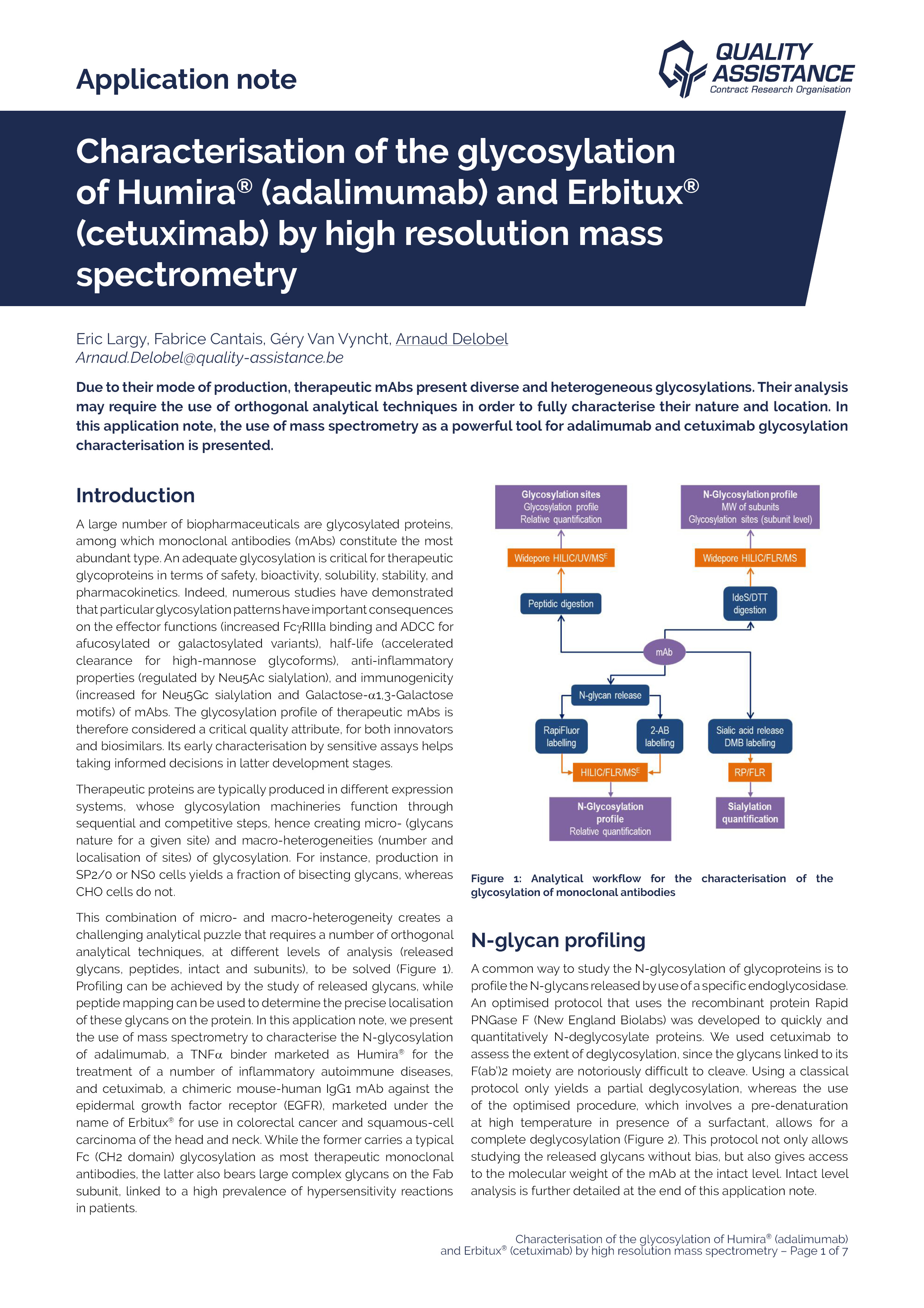
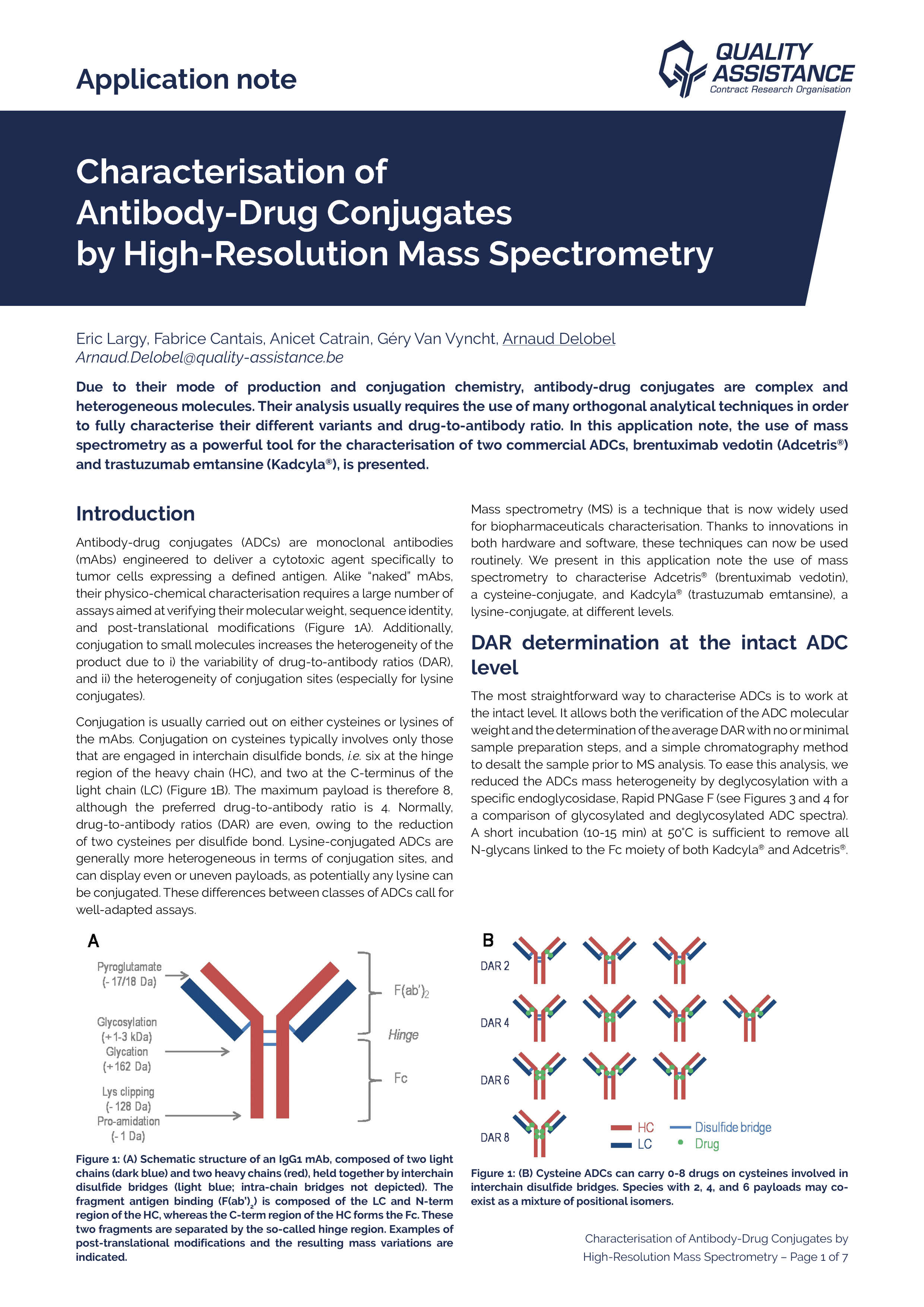
Due to their mode of production, therapeutic mAbs present diverse and heterogeneous glycosylations. Their analysis may require the use of orthogonal techniques in order to fully characterize their nature and location. In this application note, the use of mass spectrometry as a powerful tool for Adalimumab and Cetuximab glycosylation characterization is presented.

Using a comprehensive workflow combining several analytical methods at the subunit, peptide, and released glycan levels, we extensively characterised the N-glycosylation of adalimumab and cetuximab with great consistency.

Profiling of the N-glycans was performed within a day using the RapiFluor-MS labelling kit (Waters), and is fully consistent with results from the literature. The Neu5Ac and Neu5Gc sialylation was quantified by straightforward acid-induced release and DMBlabelling, starting from small amounts of mAbs. The results are consistent with the N-glycosylation profiles determined previously.

Profiling of each of the N-glycosylation sites was performed by widepore HILIC/MS, after triple enzymatic digestion, yielding short peptides and a complete sequence coverage. This stationary phase allows a complete resolution of glycosylated peptides vs. aglycosylated peptides, and MSE data provides a confirmation of
the presence of glycans. We evidenced significant differences in the glycosylation of the two cetuximab sites, the overall profile being consistent with what was found by released glycans analysis.

Analysis of mAbs subunits, generated by IdeS enzyme digestion, followed by an in situ reduction, confirmed the glycosylation profile and the localisation of the glycosylation sites, with a much easier and faster sample preparation protocol than for the peptide mapping approach. It is also a way to confirm the molecular weight of these subunits, which may be challenging because of the important heterogeneity of glycosylation and other PTMs.

This comprehensive workflow is fully applicable to potentially any mAb or ADC, using small quantities of sample, and in a reduced amount of time. Other analytical methods are available at Quality Assistance to complete the study of therapeutic mAbs glycosylation, such as relative quantification of the fucosylation, sialylation profiling by mixed-mode chromatography, and MALDI TOF profiling.

Characterisation of monoclonal antibodies by Surface Plasmon Resonance (Biacore)

Fabian Vandermeers, R&D Technical Leader

Access this 45 minute webinar to learn how Surface Plasmon Resonance can be implemented in a GMP regulated environment for the comprehensive study of monoclonal antibodies and other Biologics. 

The biological activity of monoclonal antibodies and their pharmacokinetics are highly dependent on their binding to Fcg and FcRn receptors and to their target antigen. 

An adequate control of these binding properties is therefore required for characterisation, batch release, batch-to-batch consistency and stability studies. 

Based on the example of Humira® (Adalimumab), we will present the use of Surface Plasmon Resonance technology on a Biacore T200 system for the characterisation of monoclonal antibodies as regards: 

  • Binding to Fcg and FcRn receptors 
  • Binding to target antigen (TNFa) 
  • Determination of active concentration by CFCA (Calibration-Free Concentration Analysis)
  • Comparison of sensorgrams in the context of stability or comparability studies.

All these reliable, sensitive and reproducible analyses were developed by Quality Assistance and can be used for QC and stability studies of biotech products. 

Characterisation of Anti-HCP antibodies by 2D differential Gel Electrophoresis

Arnaud Delobel
Alix Verret
Nicolas Trinchero
Géry Van Vyncht

Host-cell proteins are a major class of process-related impurities in biotherapeutics that can impact the safety and efficacy of the product. Despite the advent of advanced analytical techniques, ELISA remains as the industry gold standard for the detection and quantification of HCPs during process development and for routine testing.

However, it is necessary to understand the limitations of this technique, most of which are related to the anti-HCP antibodies used in the test. This webcast describes an alternative approach for the characterisation of the HCP coverage of these anti-HCP antibodies.

2D-DIGE is a valuable tool to evaluate the coverage of the anti-HCP antibodies. This evaluation is necessary to prove that the ELISA method that will be used for routine testing of residual HCPs in therapeutic proteins is appropriate.

Compared to traditional Western Blot approaches with visual comparisons of the gels (total proteins vs. proteins detected by the antibodies), the 2D-DIGE approach allows an easier and more reliable way of characterising the anti-HCP antibodies.

It should however be noted that even if a protein is recognised by an anti-HCP antibody on a gel, it may not be the case in ELISA, as the sandwich ELISA requires the binding of two anti-HCP antibodies per HCP molecule: this may not be possible due to insufficient tertiary space on the HCP species for antibody accessibility, or limited epitopes to bond at least two anti-HCP antibodies.

This highlights the need of orthogonal methods during early stages of development to ensure that all relevant HCPs can be correctly detected.


In addition to this application note,

Fabian Vandermeers, R&D Technical Leader, presenting this study in this video


Characterisation of Antibody-Drug Conjugates by High-Resolution Mass Spectrometry

Arnaud Delobel
Eric Largy
Fabrice Cantais
Anicet Catrain,
Géry Van Vyncht

Due to their mode of production and conjugation chemistry, antibody-drug conjugates are complex and heterogeneous molecules. Their analysis usually requires the use of many orthogonal analytical techniques in order to fully characterise their different variants and drug-to-antibody ratio. In this application note, the use of mass spectrometry as a powerful tool for the characterisation of two commercial ADCs, brentuximab vedotin (Adcetris®) and trastuzumab emtansine (Kadcyla®), is presented.

In addition to the typical LC/MS workflows aimed at mAbs characterisation, a comprehensive set of methods has been developed to characterise ADCs at the intact, subunit, and peptide level. These methods are applicable to either or both lysine- and cysteineconjugated ADCs.

Herein, determination of the drug load distribution and average DAR of Adcetris® and Kadcyla® was performed at different levels, using both MS and UV responses, with an appreciable consistency. Positional isomers of Adcetris® were distinguished by a state-of-the-art HICbased 2D-LC setup, while the position and occupancy of the numerous conjugation sites of Kadcyla® were readily determined by peptide mapping, using ETD fragmentation to relieve ambiguities at the amino-acid level.

Subscribe to